Post-Mortem: Caudal Vena Cava Thrombosis
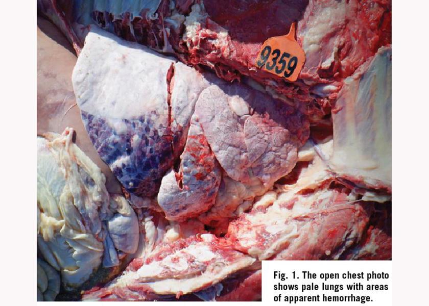
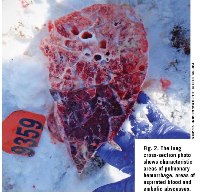
These images depict a steer calf that had been on feed for 152 days when it was found dead in the home pen.
The team diagnosed this case as a representative example of caudal vena cava thrombosis, which is a common disease of feedlot cattle, particularly later in the feeding period and in specific breeds or classes of cattle.
Pathogenesis:
Caudal vena cava thrombosis in feedlot cattle is most frequently caused by liver abscesses; however, in rare cases, caudal vena cava thromboses can also be caused by septic emboli from other diseases. Liver abscesses originate secondary to translocation of bacteria from the gastrointestinal tract to the bloodstream and ultimately the liver. Ruminal acidosis may induce rumenitis, thereby allowing for bacterial translocation.
Liver abscesses erode into the caudal vena cava and form a septic thrombus. As emboli break away from the thrombus, they often travel to the lungs and/or heart and cause embolic pneumonia and sometimes endocarditis. The emboli in the pulmonary vessels may lead to the formation of aneurisms which often rupture resulting in hemoptysis and/or epistaxis, clinical signs which characterize this disease. Sudden death, with or without clinical signs, is a common outcome in severely affected animals.
Epidemiology:
- In the feedlot, caudal vena cava thrombosis is more commonly observed later in the feeding period when cattle have been exposed to diets contain a high proportion of readily fermentable carbohydrates for a longer period of time.
- Caudal vena cava thrombosis (and liver abscessation in general) is observed more commonly in “calf-fed” dairy breeds raised for beef, but this may be due to differences in days of exposure to high-grain diets as opposed to breed differences in susceptibility.
Ante-Mortem Clinical Signs:
Animals with liver abscesses (+/-caudal vena cava thrombosis) often go undetected or are confused with other diseases until the animal has been slaughtered or succumbs to the disease. However, with respect to caudal vena cava thrombosis, secondary complications may lead to one or more of the following clinical signs:
- Bilateral epistaxis is a common clinical sign and can be foamy in appearance.
- Signs of respiratory distress may be present; including tachypnoea, dyspnoea, and coughing.
- Intermittent fever and nondescript signs of illness may be the only clinical signs in many cases.
- Melena may also be present as expectorated blood is commonly swallowed.
- Chronic cases might display weight loss and ill thrift.
Management:
Management is aimed at control and prevention of subclinical ruminal acidosis and liver abscesses. Like other metabolic diseases, a well-formulated diet with consistent bunk management, mixing, and delivery to minimize daily variations in feed intake is a key for prevention. Providing an appropriate degree of grain processing and roughage coarseness is also important. Furthermore, various in-feed antimicrobials have been proven effective at reducing the incidence and severity of liver abscesses, which in turn reduces the occurrence of caudal vena cava thrombosis.
Treatment is usually unrewarding, and prognosis is poor. If caudal vena cava thrombosis is highly suspected, humane slaughter or euthanasia may be warranted; however, a definitive diagnosis is often difficult to make with the tools available in a commercial feedlot setting.
Post-Mortem Lesions:
It is the authors’ experience that unless the caudal vena cava is thoroughly examined during the post-mortem, many cases of caudal vena cava thrombosis go undiagnosed. It would not be uncommon for the pulmonary changes observed to be confused for other types of pneumonia by the layperson. Therefore, a thorough post-mortem examination, including the caudal vena cava, is paramount to making the diagnosis. Upon post-mortem examination, lesions include:
- Pulmonary hemorrhage may result in epistaxis visible upon external examination of the carcass.
- Upon examining the open chest and cut sections of lung, any of the following lesions may be present:
- Diffuse multifocal abscesses characteristic of embolic pneumonia in a pattern indicative of hematogenous origin.
- Pallor with areas of aspirated blood and or obvious aneurysm (Figs. 1 and 2).
- Non-descript interstitial pneumonia pattern or “reactive” appearance.
- Some cases may have minimal or no lung lesions.
- Upon examining the open abdomen, hepatomegaly and/or ascites may be present if the thrombus has sufficiently occluded the vena cava.
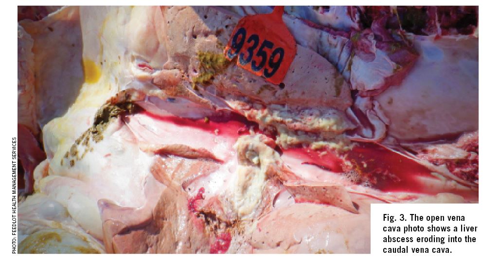
- Upon incising the liver and examining the vena cava, the pathognomonic lesion consists of an abscess protruding into the vena cava and presence of a septic thrombus (Fig. 3). Additionally, passive congestion of the liver grossly observed as “nutmeg” liver may also be apparent if the vena cava has been occluded.
For more information about Feedlot Health Management Services, visit their website at www.feedlothealth.com


