Post-Mortem: Ruminal Tympany Or Bloat in the Feedlot?
This article is from our Post-Mortem Series that was done in partnership with Feedlot Health Management Services, Okotoks, Alberta.
These images depict a steer calf that had been on feed for 272 days with no treatment history when it was found dead in the pen.
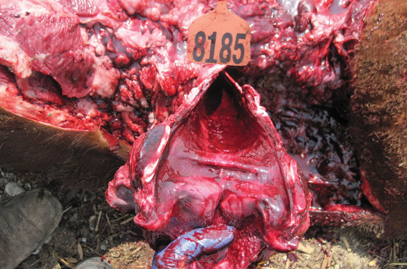
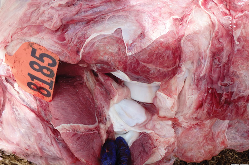
The team diagnosed this case as a representative example of ruminal tympany or bloat, which is a common disease of feedlot cattle, particularly later in the feeding period.
Pathogenesis:
Bloat is a metabolic syndrome caused by an imbalance between the volume of gas produced in the rumen and the volume of gas released via normal eructation, with the scales tipped in the direction of the former. This imbalance can be caused by physical impairment of the ability to eructate, entrapment of gas in a foam or slime, and/or rapid fermentation of grain associated with things such as: inconsistent feed intake, ration step-ups, feed mixing errors, or too fine of grain particle size (especially with more rapidly fermentable small grains such as wheat or barley). Accumulated gas from fermentation distends the rumen and greatly increases intra- abdominal pressure. Consequently, the thorax becomes compressed while caudal circulation is impeded, resulting in dyspnea. Without timely treatment, death by asphyxiation is common.
Epidemiology:
- In the feedlot, bloat is more commonly observed later in the feed period when diets contain a high proportion of readily fermentable carbohydrates. As cattle are fed high concentrate diets, ruminal motility may also be decreased. The risk for bloat and other metabolic issues increase as cattle are fed high concentrate diets, this risk increases the longer the cattle are fed these diets.
- Bloat may also occur early in the feeding period but this is commonly associated with other co-morbidities that occur during this time.
- Bloat may occur secondary to ruminal acidosis, due to decreased rumen motility and therefore decreased ability to eructate.
- Bloat can also be a chronic, recurrent condition caused by damage to either the rumen itself or the vagus nerve, which controls rumen motility. The vagus nerve can be compressed or damaged by chronic pneumonia, pleuritis, or esophageal obstructions/lesions.
- Sporadic cases of bloat may occur at any time if the animal is unable to eructate due to recumbency (e.g., if the animal is cast).
Ante-Mortem Clinical Signs:
- The abdomen becomes dorsal-laterally distended with the left paralumbar fossa more obviously affected.
- As the abdominal pressure increases, animals become dyspneic, with an extended neck and open mouth breathing.
- Bloated animals become reluctant to move and are often recumbent as the dyspnea and hypoxia progress. Animals may also become increasingly agitated.
Management:
Consistency is key for bloat prevention, and management involves providing a well-formulated diet with consistent feed calling, mixing, and delivery. Grain to roughage ratio is an important consideration, as well as the coarseness and degree of processing of the grain and roughage. Some supplements, such as ionophores, can also reduce the risk of bloat through regulation of feed intake and alteration of the rumen microenvironment.
The primary treatment objective is to relieve the accumulated rumen gas and avoid asphyxiation. In many cases, simply inserting a stomach tube is enough. If the animal is recumbent or in immediate risk of asphyxiation, emergency rumen trocarisation can be a necessary life saving procedure. Animals who repeatedly bloat (possibly due to vagal indigestion) can be treated surgically by performing a rumenostomy.
Post-Mortem Lesions:
- It is important to note that although a carcass may have an obviously distended rumen causing separation of the hind limbs, other pathology must be observed to confirm free gas bloat as the cause of death. Many carcasses will “bloat” as part of post-mortem autolysis, especially in warmer climates, making a full post-mortem examination paramount to diagnosing bloat mortalities. Many feedlot personnel will incorrectly diagnose animals as bloat based on external appearance alone.
- Upon examining the open chest and abdomen, the lungs and liver are significantly compressed and dorsally displaced. Most commonly, the lungs are slightly to moderately congested, while the liver and heart are pale (Fig. 1).
- Cranial congestion is specifically evident in hemorrhagic trachea mucosa and cervical tissues (Fig. 2).
- Along with caudal pallor, subcutaneous edema and/or emphysema is often present in the hindlimb musculature (Fig. 3).
For more information about Feedlot Health Management Services, visit their website at www.feedlothealth.com.


