Post-Mortem of a Heifer on Feed: Answer


The team diagnosed this case as “Myocarditis” most likely caused by Histophilus somni, a component of the bovine respiratory disease (BRD) complex; however, the septicemic form can cause myocarditis, pericarditis, pleuritis, arthritis, and infectious thromboembolic meningoencephalitis.
Pathogenesis
- Generally considered a normal inhabitant of the nasopharynx that gains access to the bloodstream (potentially due to immune stressors and/or respiratory tract infection)
- Predilection for vascular endothelium leads to adherence and thrombus formation followed by ischemia, necrosis, and eventually sequestration in specific organs.
Epidemiology
- Most commonly observed in calves placed in late fall and early winter, but the seasonal effect is confounded by the high number of calf placements in these seasons
- Mortalities caused by Histophilus somni predominately occur between 30 and 60 DOF
Ante-Mortem Clinical Signs
- Animals may exhibit non-specific clinical signs such as depressed mentation, fever, anorexia, and/or lethargy
- In the acute stage of infection, it is difficult to clinically differentiate between H. somni caused myocarditis and BRD; therefore, ante-mortem cases of both disease syndromes are most accurately categorized as “undifferentiated fever”
- Compared to BRD, H. somni caused myocarditis is approximately 2 times more likely to be missed by stock attendants and animals are commonly found dead in the pen without prior treatment history
Management
- In populations at high risk of BRD, metaphylaxis with tulathromycin has been shown to reduce H. somni specific mortality.
- Vaccination with an H. somni bacterin has been evaluated with mixed results; however, studies have generally used commingled experimental designs with vaccinates and non-vaccinates in the same pens
- In an outbreak scenario, H. somni is generally susceptible to tetracycline antimicrobials. Due to the difficult nature of identifying H. somni caused disease, mass medication may be warranted.
Post-Mortem Lesions
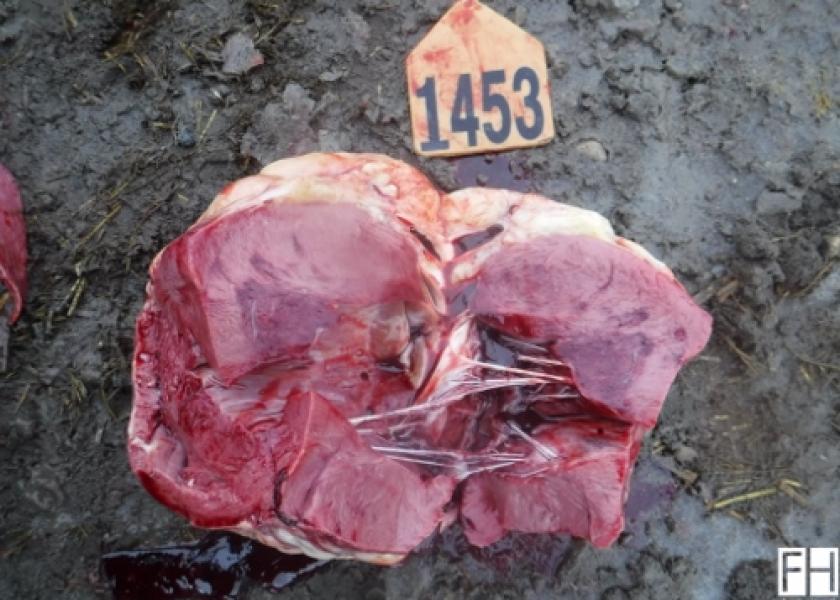
- Upon opening the left ventricle of the heart and incising the papillary muscles, acute myocardial necrosis appears as a circumscribed area that is darker in color (purple) than the surrounding tissue located within the papillary muscles (see Fig. 1).
- If the animal survives the acute phase of disease, a sequestra often forms in the papillary muscles of the left ventricle. Figure 2 shows the open left ventricle of a steer calf that died at 77 DOF and had previously been treated twice for undifferentiated fever.
- Upon observation of the open chest, cardiogenic pulmonary edema may be apparent with all stages of disease; however, it is most commonly observed in later stages of the disease (Fig. 3).
Post-Mortem Series presented in partnership with Feedlot Health management Services, Okotoks, Alberta. For more information, visit their website at www.feedlothealth.com.
Working with crews at client operations, Feedlot Health conducts post-mortem exams on all feedlot and calf-grower mortalities, using a standard protocol for recording the animal’s history, digital images, and post-mortem findings. The group compiles images and post-mortem findings in a central database, for review by the professional team, as an educational tool and to track disease trends within an operation or across their client base.